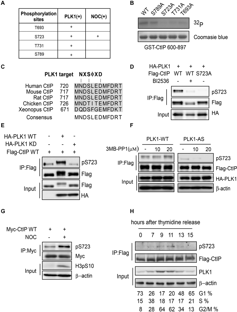
Figure 3.
CtIP is phosphorylated by PLK1. (A) CtIP phosphorylation by PLK1 was analyzed by mass spectrometry and the potential PLK1 target sites on CtIP were identified. PLK (+): purified CtIP protein was incubated with PLK1 in vitro and then subjected to mass spectrometry. NOC (+): U2OS cells stably expressing Flag-CtIP were incubated with nocodazole (NOC) for 16 h before collection. Flag-CtIP protein was immunopurified from the cell lysate using an anti-Flag antibody and subjected to mass spectrometry. (B) Purified GST-CtIP fragments (600-897) and the indicated mutants were incubated with [γ-32P] ATP in the presence of PLK1 for the in vitro kinase assay. The radio-labeled proteins were visualized following SDS-PAGE. Coomassie Blue staining indicates the protein loading. (C) Sequence alignment of the CtIP S723 surrounding region, showing a canonical PLK1 target sequence and the PLK1 target residue S723. (D) 293T cells were co-transfected with Flag-tagged CtIP and HA-tagged PLK1 expression constructs, and treated with or without 10 μM BI2536 for 1 h before harvesting. Cell extracts were subjected to immunoprecipitation followed by western blotting with the indicated antibodies. (E) 293T cells were co-transfected with Myc-tagged CtIP and Flag-tagged PLK1 WT or PLK1-KD (kinase dead K82M/D176N) expression constructs. Cell extracts were subjected to immunoprecipitation followed by western blotting with the indicated antibodies. (F) 293T cells were co-transfected with Flag-tagged CtIP and HA-tagged PLK1 WT or analog-sensitive PLK1 mutant (PLK1-AS) expression constructs, and treated with or without the indicated amounts of 3-MB-PP1 for 16 h. The cell extracts were subjected to immunoprecipitation followed by western blotting with the indicated antibodies. (G) U2OS cells stably expressing Myc-tagged CtIP were treated with nocodazole for 16 h before harvesting. Cell extracts were subjected to immunoprecipitation followed by western blotting with the indicated antibodies. (H) CtIP-knockout (KO) HCT116 cells stably expressing Flag-CtIP WT were synchronized by double thymidine-block release; CtIP S723 phosphorylation was analyzed using pS723 antibody. Cell-cycle progression was analyzed by FACS.

