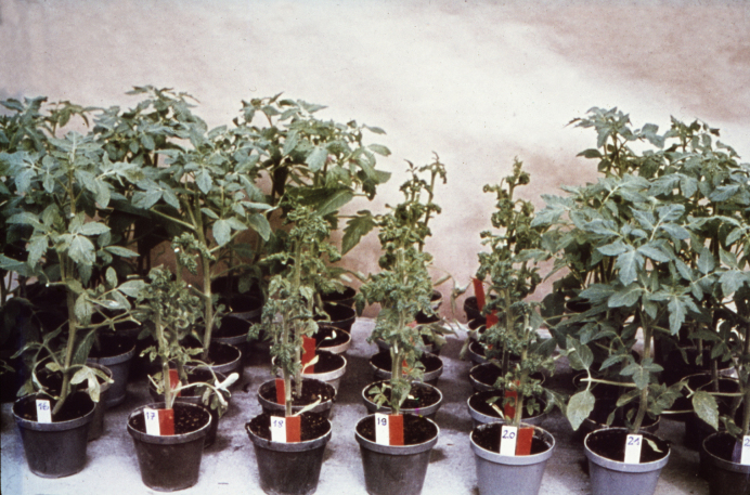
Figure 3.
An early assay method for viroid. A crude RNA extract from a PSTVd-infected tomato plant was separated by denaturing PAGE (cf. lane ‘M’ of Figure 5B). After electrophoresis the gel under the slot of application was cut horizontally into about 30 slices, the RNA eluted and a series of tomato plants inoculated. The photograph shows a ‘tomatogram’, in which the front row of tomato plants was inoculated with a higher concentration of RNA than the back row. Clearly, the highest concentration of PSTVd RNA was in gel slices 19 and 20. The photograph is courtesy of Heinz-Ludwig Sänger(†), Martinsried.

