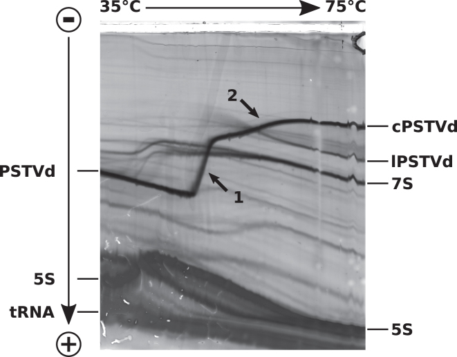
Figure 9.
Temperature gradient gel electrophoresis. TGGE analysis of a crude RNA extract from tomato plants infected with PSTVd. A linear temperature gradient is applied perpendicular to the electric field. The sample is loaded into the broad central slot (top). RNAs were visualized by silver staining. The rod-like structure of circular PSTVd (cPSTVd; 359 nt) migrates relatively fast at low temperature; it denatures in a highly cooperative transition (see arrow 1) into a mostly single-stranded structure with at least two extrastable hairpins I and II; these hairpins denature above the main transition (see arrow 2). Linear PSTVd (lPSTVd) molecules migrate proportional to their length after full denaturation and faster than fully denatured cPSTVd. The temperature of the main transition of lPSTVd depends on the site of linearization; lPSTVd is not visible at low temperatures due the low concentration of individual lPSTVd molecules with different 5′- and 3′-ends. The different 7 S RNAs (∼305 nt) are separated at low temperature due to their different thermodynamic stability but co-migrate at high temperature due to their nearly identical length. 5S: 5 S ribosomal RNA (120 nt).

