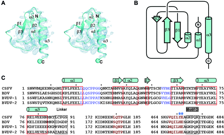
Figure 2.
The structure, folding topology and sequence analysis of the CSFV NS5B NTD. (A) Stereo-pair images of the NTD with 3500 K composite SA-omit electron density map (contoured at 1.2σ) overlaid. (B) Topology of the CSFV NS5B NTD. The α-helix and β-strand labeling in panels A and B is based on secondary structure assignment by DSSP (68). (C) The sequence alignment of the pestiviruses NS5B NTD, linker and the RdRP regions that interact with NTD. The symbols of NTD α-helices (cylinders) and β-strands (block arrows) are plotted based on the secondary structure assignment of the CSFV NS5B structure. Two key residues involved in the intra-molecular of NTD–RdRP interactions and chosen for mutagenesis are indicated by asterisks. UniProtKB accession numbers of the pestivirus NS5B sequences used: Q5U8X5 (CSFV), X2KMP2 (BDV), P19711 (BVDV-1) and A0A0M4S9G5 (BVDV-2).

