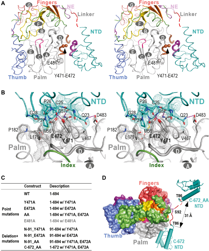
Figure 3.
The intra-molecular NTD–RdRP interface and an open-conformation NS5B structure derived from the interface mutations. (A) Stereo-pair images of CSFV NS5B structures viewing from the NTP entry channel. The seven CSFV RdRP catalytic motifs A-G are labeled in the structures, and motifs A and D are shown as thick noodles. The key regions from NTD and RdRP that form the NTD–RdRP interface are colored in purple and brown, respectively, while all other regions are colored as in Figure 1A. The α-carbon atoms of residues 471, 472 and 481 are shown as spheres. Red dotted line in the structure indicates residues 87–88 that were not modeled due to poorly defined electron density. (B) Stereo-pair images of the NTD–RdRP interface with 3500 K composite SA-omit electron density map (contoured at 1.5σ) in the vicinity of the interface overlaid. Side chains of residues 471–472 (thick) and other residues (thin) that participate in the interface interactions are shown in sticks. The hydrogen bonds are shown as purple dotted lines. (C) A list of NS5B constructs used to investigate the structure and function of NTD, with full descriptions including residue range, mutation site and mutation type. (D) A comparison of the global conformations observed in the crystal structures of the CSFV NS5B C-672 and the interface mutant C-672_AA. NTD and linker are shown in noodle/cylinder style, and RdRP is shown as surface representation. The α-carbon atoms of residues T86 and S92 adjacent to the NTD–RdRP linker are shown as spheres. The coloring scheme is as in Figure 1A.

