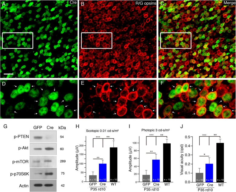
Fig. 3. The conditional deletion of PTEN in cone photoreceptors of rd10 mice in vivo.
a–f Flat-mounted retinas of rd10/PTENloxP/loxP mice treated with AAV-hRo (human red opsin promoter)-Cre and harvested at P25. Representative retinal flat mounts show extensive Cre expression stained by an anti-Cre antibody (a, green) and red/green cones labeled by red/green opsins (b, red). The majority of cre-positive cells were also positive for red/green opsins (c). d–f Illustrate highly magnified images from the boxed regions above, respectively. Arrows indicate the colocalization of Red/green cones with Cre stain. Scale bar: 20μm. g Western blots of whole retinal p-PTEN and t-PTEN, p-Akt and t-Akt, p-mTOR and t-mTOR, and p-p70S6K from Cre- or GFP-treated PTENloxP/loxP/rd10 mouse retinas. β-actin levels were used as a loading control. h Average scotopic b-wave amplitudes from P35 AAV-Cre- (blue bar) or AAV-GFP-treated PTENloxP/loxP/rd10 mice (gray bar). Age-matched C57BL/6J are shown as comparisons (black bar). i Averaged photopic b-wave amplitudes from the three groups of mice. j Photopic visual acuity was measured by optokinetic responses. Results are presented as the mean±SD (n = 10). *p < 0.05, **p < 0.01, ***p < 0.001

