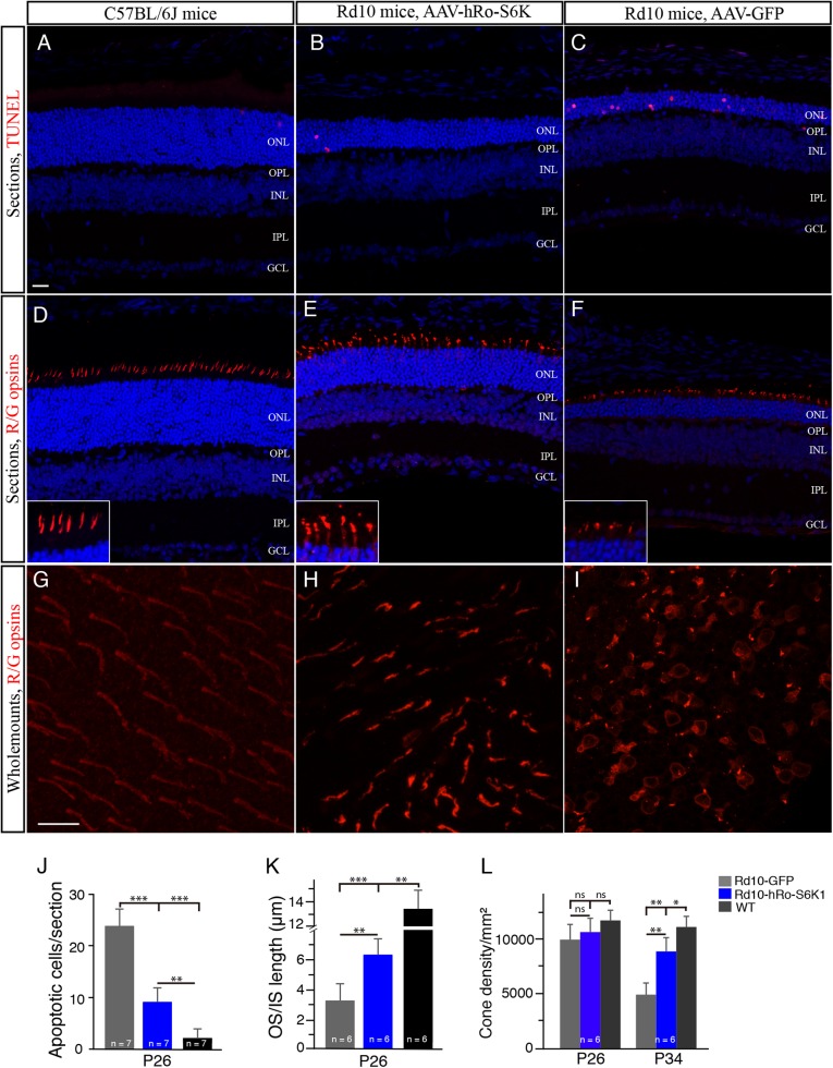
Fig. 6. S6K1 overexpression in cones driven by a hRo promoter enhances the survival of the cones in rd10 mice.
a–c Retinal sections from S6K1- and GFP-treated P26 rd10 mice and WT mice were stained with TUNEL (red). The nuclear layers in retinal sections were stained with diamidinophenylindole (DAPI, blue). d–f Retinal sections from S6K1- and GFP-treated P26 rd10 mice and WT mice were stained with an antibody against red/green opsin (red). The nuclear layers in retinal sections were stained with DAPI (blue). Insets are highly magnified images from respective images showing cone OS/ISs. ONL outer nuclear layer, OPL outer plexiform layer, INL inner nuclear layer, IPL inner plexiform layer, GCL ganglion cell layer, OS outer segment, IS inner segment. Scale bar: 20μm. g–i Confocal images of red/green cones in the dorsal retina of retinal flat mounts, 1mm superior to the center of the optic nerve are shown. Retinal flat mounts from P34 rd10 mice treated with S6K1 or GFP and age-matched WT mice were stained with an antibody against red/green opsins. Scale bar: 20μm. j Quantification of TUNEL-positive photoreceptors from retinal sections. k Plot of the length of cone OS/ISs measured on retinal sections. l Quantification of red/green cone density in the dorsal retina of retinal flat mounts. Results are presented as the mean±SD. ns, not significant, *p < 0.05, **p < 0.001, ***p < 0.0001

