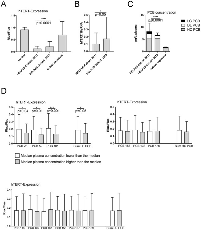
Figure 1.
Inhibition of hTERT gene expression by blood plasma of PCB exposed individuals. (A) Tert+ B6B5.1 cells were incubated with longitudinally collected plasma samples from the HELPcB-cohort (N = 92 per time point; each with three technical replicates) or with plasma samples from individuals exposed to PCB via indoor air (N = 105; each with three technical replicates). After 48 hours of incubation, luciferase activities were determined. Statistical analysis was performed by a Wilcoxon matched-pairs signed rank test. Statistically significant differences are indicated (*P = 0.05; ****P < 0.0001). (B) PBMCs from healthy donors (0 RhD negative) were stimulated with tetanus toxoid for five days, reseeded and incubated in the presence of antigen- and PCB-containing plasma samples from the HELPcB-cohort as described in A. After 48 hours of incubation, hTERT gene expression was assessed by qRT-PCR. Statistical analysis was performed by a Wilcoxon matched-pairs signed rank test. (C) Mean plasma levels for Σ higher chlorinated PCBs (grey), Σ dioxin-like PCBs (white) and Σ lower chlorinated PCBs (black) from longitudinally collected samples (HELPcB-cohort) and from individuals exposed to PCB via indoor air. Statistical analysis was performed by a Wilcoxon matched-pairs signed rank test. SD and statistically significant differences are given for Σ lower chlorinated PCBs (****P < 0.0001). (D) Mean ± SD ratio of hTERT expression (N = 184) for individual indicator PCBs depending on plasma concentration. HC PCBs, DL PCBs and LC PCBs were separated using the median value as cut off. Statistical analysis was performed by a Mann-Whitney test. Statistically significant differences are indicated.

