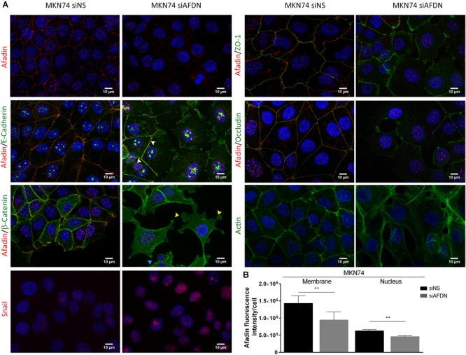
FIGURE 3.

Afadin downregulation displaces apical junctional complex proteins and promotes the formation of actin stress fibers. Double immunofluorescence of Afadin (red) with AJs proteins E-cadherin and β-catenin (green), and with TJs proteins ZO-1 and occludin (green) in MKN74 cells transfected with a non-silencing siRNA (siNS) or with a siRNA to Afadin (siAFDN). Immunofluorescence of actin (green) and of Snail (red) are also shown. Nuclei were counterstained with DAPI. Scale bar, 10 μm. White arrows represent cells with Afadin not efficiently silenced by the siRNA and that retain the epithelial morphology; Yellow arrows, lamellipodia; cyan arrows, filopodia (A). Quantification of Afadin fluorescence intensity in MKN74 cells in both membrane and the nucleus upon treatment with non-silencing siRNA or with a siRNA to Afadin (B).
