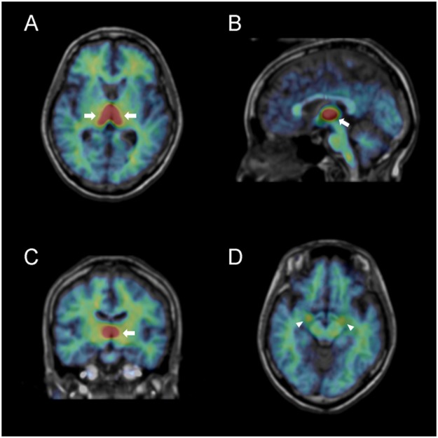
Figure 1.
Representative distribution volume (BPND + 1) images of 11C-cetrozole in a female human brain (rainbow colour scale) superimposed on the structural MR image (gray scale) of the same subject. (A) transaxial slice at level of the thalamus; (B) sagittal slice at the midline; (C) coronal slice at the level of the thalamus; (D) transaxial slice at the level of the amygdala. Arrows and arrow heads indicate the thalamus and amygdala, respectively.

