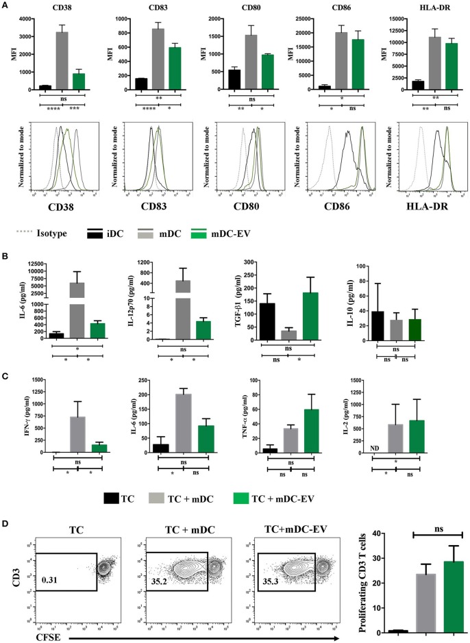
Figure 4.
MSC-EVs modulate DC maturation and cytokine secretion. (A) Cumulative (top) and representative (bottom) data showing expression of surface markers CD38, CD83, CD80, CD86 and HLA-DR as determined by flow cytometry on immature, mature and MSC-EV treated mature DCs. (B) Cell culture supernatants collected from iDC, mDC and mDC-EVs were analyzed for the levels of IL-6, IL-12p70, TGF-β and IL-10. (C) Supernatants collected from the co-culture of CD3+ T cells with either mDCs or mDCs-EVs were analyzed for the levels of IFN-γ, IL-6, TNF-α and IL-2. T cells cultured with medium alone served as a control. (D) Representative (left) and cumulative (right) data showing allogeneic CD3+ T cell proliferation stimulated with mDCs and mDC-EVs as assessed by flow cytometry analysis of CFSE dilution. Data represent mean ± SEM of 4–6 independent experiments. *p < 0.05, **p < 0.01, ***p < 0.001, ****p < 0.0001. ND, not detectable.

