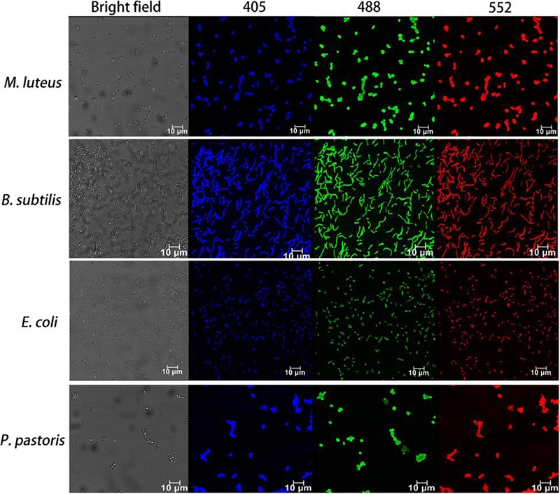
FIGURE 6.

Fluorescence images of dead microorganisms covering two other Gram-positive bacteria (M. luteus and B. subtilis), one Gram-negative bacterium (E. coli) and yeast (P. pastoris) labeled with CDs-EPS605. The corresponding bright field was also presented. Fluorescence images were observed with the excitation length of 405, 488, and 552 nm, respectively.
