
Figure 2.
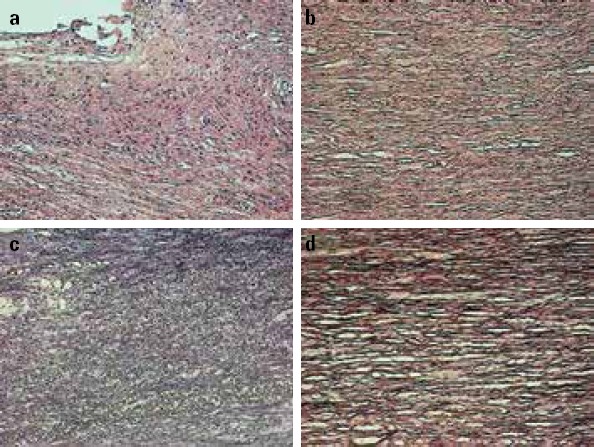
Pathomorphological observations of the aortic vessel specimens of the aortic dissection (AD) and aortic aneurysm (AA) groups using hematoxylin–eosin (HE) and elastic fiber (EF) staining technique under 200× magnification
(a) HE staining of the specimen in the AD group
(b) HE staining of the specimen in the AA group
(c) EF staining of the specimen in the AD group
(d) EF staining of the specimen in the AA group