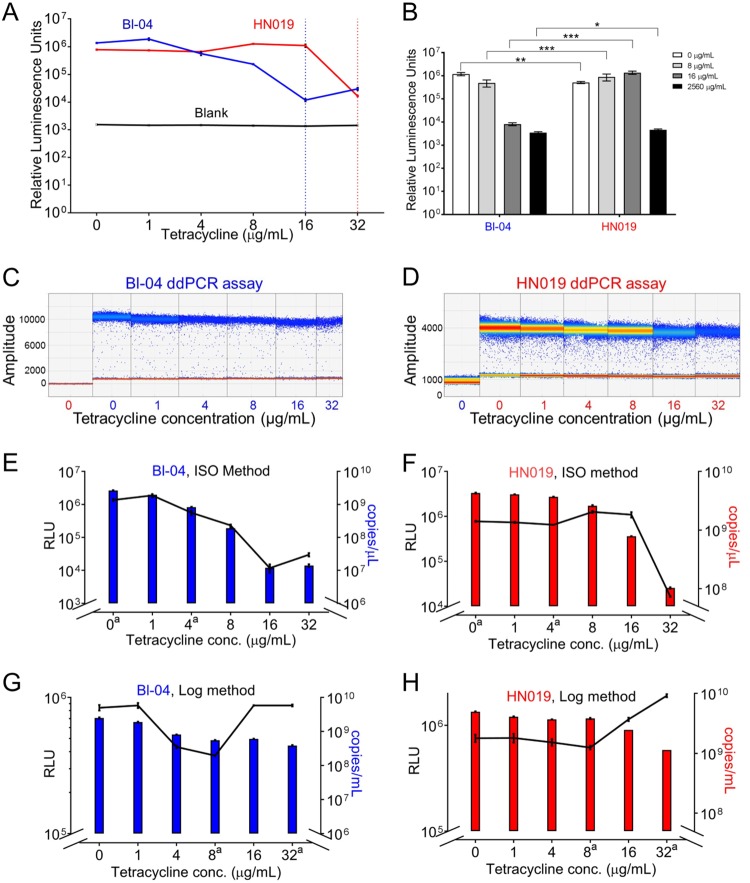
FIG 2.
Assay of tetracycline response using ATP luminescence and droplet digital PCR. (A) ATP quantification using the microdilution procedure, with the dotted vertical lines showing the MIC levels for both strains. (B) The difference in ATP response between the two strains at the MIC thresholds (*, P < 0.01; **, P < 0.001; ***, P < 0.0001; paired t test). Droplet digital PCR results for the (C) Bl-04 and (D) HN019 assays show number of droplets per amplitude with a quantitative heat map applied. Sample colors in the x axes denote Bl-04 (blue) and HN019 (red) by tetracycline concentration. Droplet digital PCR assay results are shown as colored bars for (E) Bl-04 and (F) HN019 compared to ATP concentration (black lines). The experiment was repeated using acute exposure to tetracycline for (G) Bl-04 and (H) HN019. The y axes have been adjusted to better show overall correlation. aConditions used for the RNA-seq experiments.

