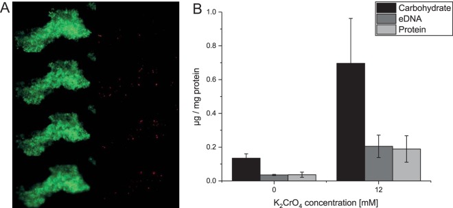
FIG 2.
Chromate stress-induced aggregate formation of L. chromiiresistens. (A) LIVE/DEAD stain of a representative aggregate. Green, 4′,6-diamidino-2-phenylindole (DAPI) stain (all cells); red, propidium iodide stain (cells with collapsed membrane potential). Shown are 4 different Z-levels of the aggregate. (B) EPS components of cellular aggregates of L. chromiiresistens cells grown without (0 mM) and with (12 mM) K2CrO4. The quantity of each of the tested components increased as a response to growth under chromate stress. All values were normalized to the initial amount of biomass (quantified as total cell protein) that was used for EPS isolation. Error bars represent SD of the results from three independent replicates.

