FIG 4.
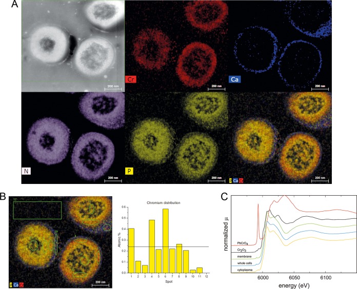
Chromium distribution in L. chromiiresistens extracellular matrix. (A) EDS images of chromate-grown cells. STEM overview (HAADF) and element distribution. Cr, chromium; Ca, calcium; N, nitrogen; P, phosphorus; PCaCr, superimposition of P, Ca, and Cr signals. (B) Detailed element analysis of the chromium dispersion. The spots are indicated on the left, and the corresponding atomic percentages of chromium are shown on the right. The black line indicates the mean chromium content of all analyzed spots (1 to 11). (C) Determination of chromium oxidation status via XANES. The predominant chromium species bound to cells was identified as Cr3+.

