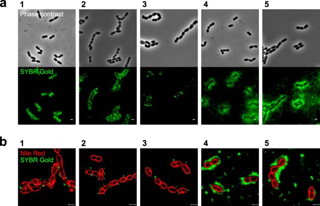
FIG 2.
Fluorescence imaging of phage binding to S. thermophilus strains. (a) Adsorption of phages to their hosts was visualized with a conventional fluorescence microscope after labeling phage DNA with SYBR Gold. (b) Superresolution structured illumination microscopy (SR-SIM) images of bacterial cells stained with Nile red (red) and mixed with SYBR Gold DNA-labeled phages (green). Panels with phages and their host strains: 1, CHPC926 and STCH_15; 2, CHPC951 and STCH_12; 3, CHPC1057 and STCH_09; 4, CHPC1014 and STCH_13; 5, CHPC1046 and STCH_14. Two binding patterns are observed: spotty (panel numbers 1, 2, and 3) or diffused (panel numbers 4 and 5). Scale bars, 1 μm.

