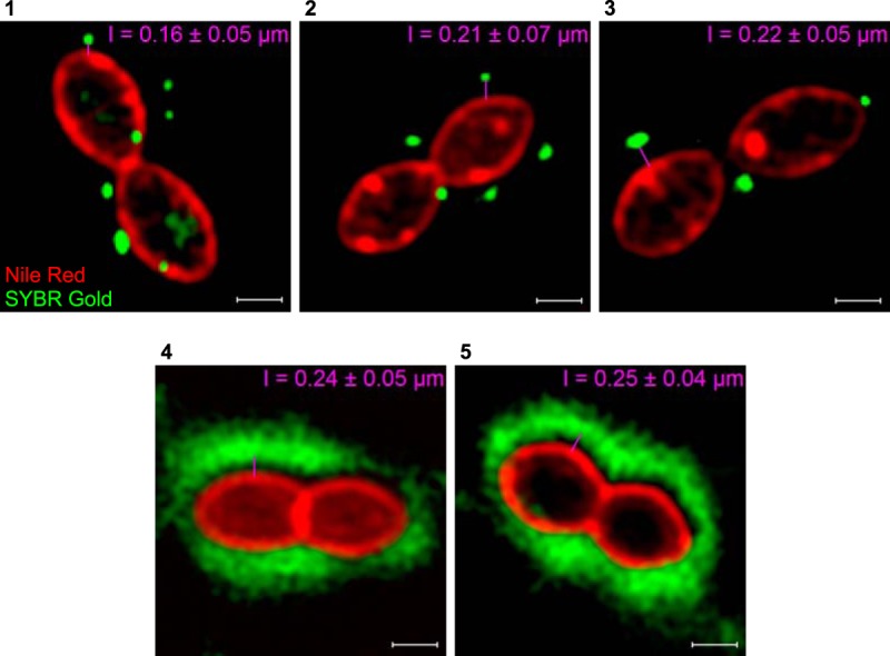
FIG 3.

Superresolution structured illumination microscopy (SR-SIM) images of phage binding to S. thermophilus strains. The pink lines indicate the distance between phage capsids, containing SYBR Gold-labeled DNA (green), and bacterial membranes, stained with Nile red (red). The presented values correspond to the lengths of phage tails and are the averages from 80 measurements. Panels with phages and their host strains: 1, CHPC926 and STCH_15; 2, CHPC951 and STCH_12; 3, CHPC1057 and STCH_09; 4, CHPC1014 and STCH_13; 5, CHPC1046 and STCH_14. Scale bars, 0.5 μm.
