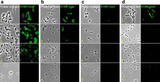
FIG 4.
Fluorescence imaging of phage binding to cellular fractions of S. thermophilus. Phage DNA was labeled with SYBR Gold and the green fluorescence was visualized. (a) Phage CHPC951 was mixed with cellular fractions of STCH_12. Phage CHPC926 was mixed with cellular fractions of STCH_15 (b), cellular fractions of STCH_15_BIM_1 (c), or cellular fractions of STCH_15_BIM_2 (d). The following samples were used: 1, cells in exponential phase; 2, cells devoid of surface enzymes, membranes, and membrane proteins; 3, purified cell walls; 4, purified peptidoglycan. Scale bars, 1 μm.

