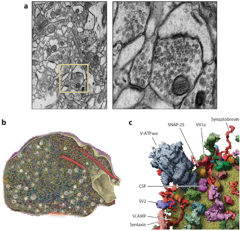
Figure 1.
Presynaptic terminals are complex structures. (a) Transmission electron micrograph taken from rat hippocampal neuropil highlighting presynaptic terminals. The right panel is a close-up view of the yellow box in the left panel. Modified from Atlas of Ultrastructural Neurocytology on Synapse Web by Josef Spacek, Kristen Harris, and John Fiala. http://synapseweb.clm.utexas.edu/atlas. (b) Quantitative three-dimensional model of a section through an average presynaptic terminal displaying >300,000 proteins in atomic detail. The active zone is shaded red at bottom. From Wilhelm et al. (2014). (c) Three-dimensional model of a prototypical synaptic vesicle (close up) containing dozens of proteins in stoichiometrically accurate atomic detail. From Takamori et al. (2006).

