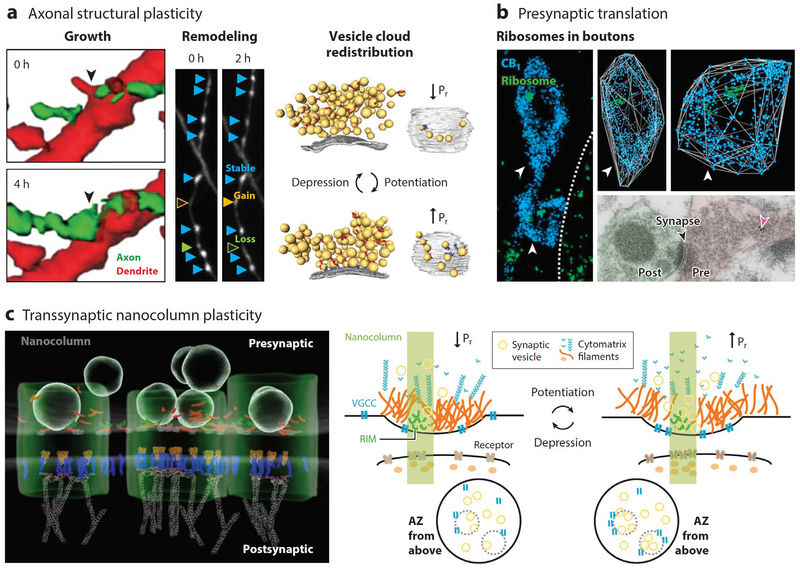
Figure 2.
Emerging molecular mechanisms of presynaptic plasticity. (a) Different forms of axonal structural plasticity. (Left) Growth: Inhibitory axon (green) form a contact on a hippocampal pyramidal cell dendrite (red). Arrow indicates the location of a future bouton. Modified from Wierenga et al. (2008). (Middle) Remodeling: GABAergic boutons can be stable (light-blue arrowheads), gained (yellow arrowheads), or lost (orange arrowheads). Modified from Schuemann et al. (2013). Images collected from organotypic hippocampal slice cultures using time-lapse, two-photon microscopy. (Right) Vesicle cloud redistribution: Synaptic vesicle (yellow) number, density, proximity to, and priming or docking state at the AZ(gray) can contribute to Pr. Cytomatrix filaments (red and blue) may also help regulate Pr. Modified from Fernández-Busnadiego et al. (2013). Data collected from cerebrocortical synaptosomes by way of cryo-electron microscopy. (b) Ribosomes in presynaptic boutons. (Left and top-right) Dual-STORM imaging of CB1 receptors delineating two GABAergic boutons (blue) and 5.8S ribosomal rRNA (green) in acute hippocampal slices. Three-dimensional (3D) reconstructed boutons shown at right. Arrowheads indicate the two boutons. Dotted line in left panel outlines CA1 pyramidal cell soma. From Younts et al. (2016). (Bottom-right) presynaptic ribosomes (pink arrow) revealed by immunogold electron microscopy on retinal ganglion cell axon in the superior colliculus. Modified from Shigeoka et al. (2016). (c) (Left) Model of synaptic nanocolumn based on data collected by 3D-STORM revealing alignment of presynaptic and postsynaptic molecules. Shown presynaptically are synaptic vesicles, AZ proteins RIM1/2 (red) and Munc13 (green). Shown postsynaptically are glutamate receptors (orange), PSD-95 (blue), and actin (gray). The green tube highlights the nanocolumn. Modified from Tang et al. (2016). (Right) Schematic representation of the nanocolumn and its modulation. Plasticity can result from changes in presynaptic vesicle or cytomatrix filament clustering or changes in the density or proximity of VGCCs to the active zone. Adapted from Glebov et al. (2017). Abbreviations: AZ, active zone; Pr, probability of release; PSD, postsynaptic density; RIM, Rab-interacting proteins; STORM, Stochastic optical reconstruction microscopy; VGCC, voltage-gated Ca2+ channel.

