Figure 1.

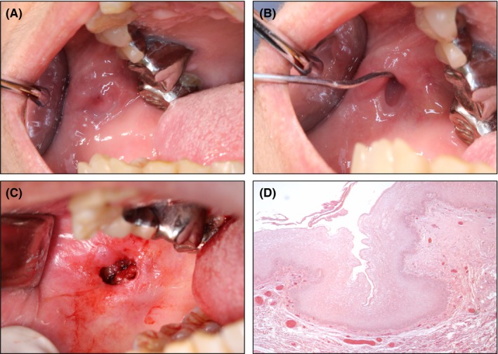
Intraoral view and histological image. A, Intraoral view at the first visit. B, Intraoral view showing the diverticular orifice. C, Intraoperative view. D, Histological image

Intraoral view and histological image. A, Intraoral view at the first visit. B, Intraoral view showing the diverticular orifice. C, Intraoperative view. D, Histological image