Fig. 3.

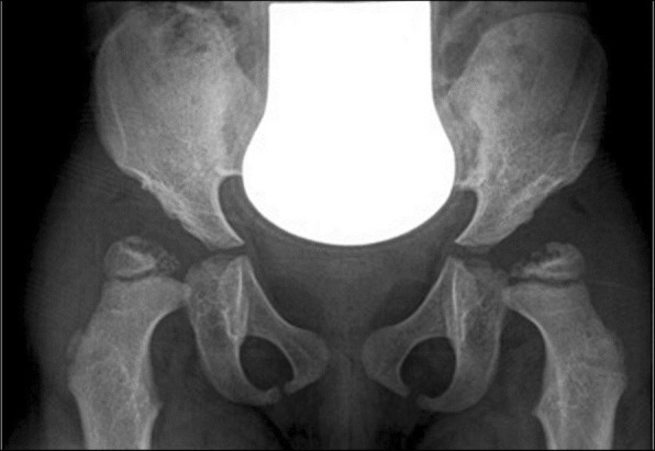
4-year-old with MPS VI. X-ray of the pelvis showing enlarged and receding acetabulum, underdeveloped femoral epiphysis, and acetabulum warped with coxa valga

4-year-old with MPS VI. X-ray of the pelvis showing enlarged and receding acetabulum, underdeveloped femoral epiphysis, and acetabulum warped with coxa valga