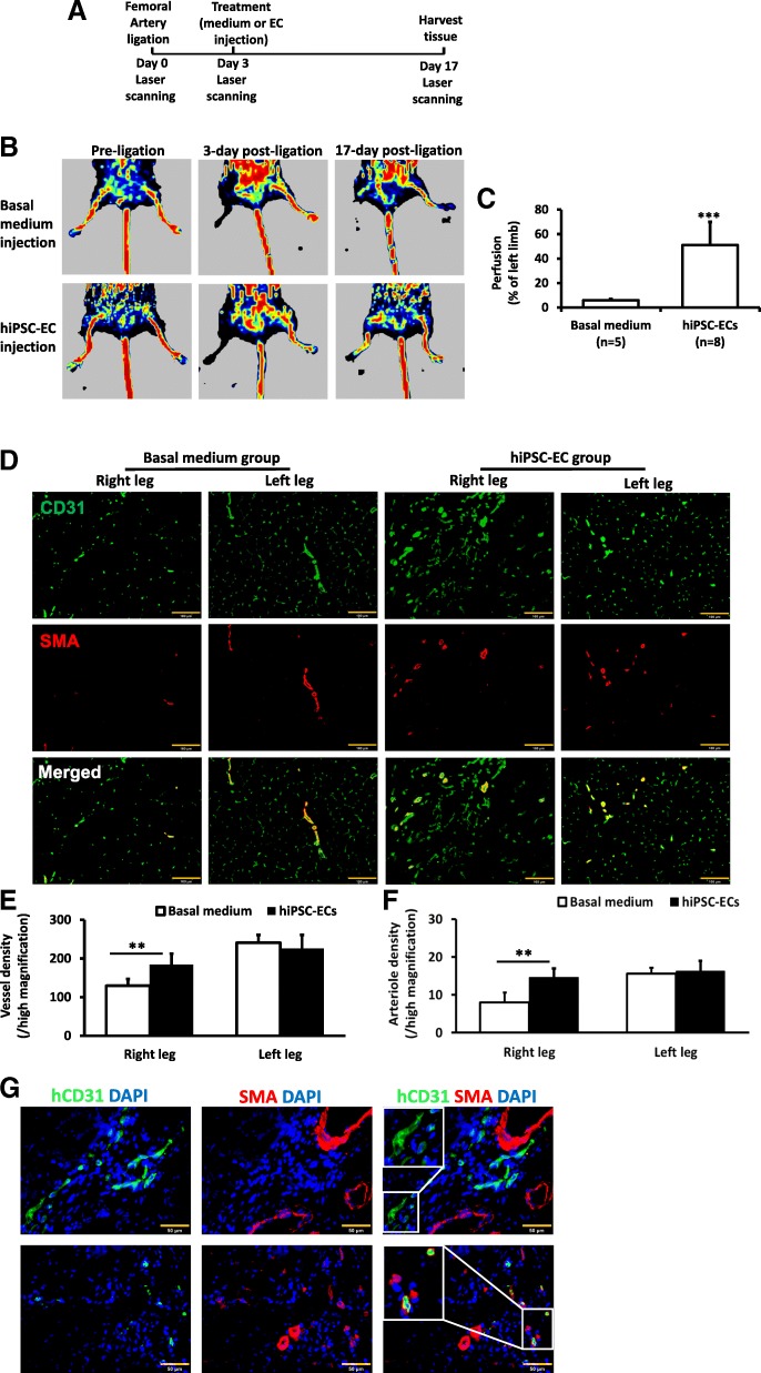
Fig. 6.
a A schematic diagram of the HLI model and treatment. b Laser Doppler imaging of mouse limbs before femoral artery ligation, 3 days after femoral artery ligation (i.e., at the time of treatment administration) and 17 days after treatment with basal medium or hiPSC-ECs. Laser Doppler imaging was performed with a PeriScan PIM 3 System with a similar setting. c Recovery of right limb perfusion was expressed as a percentage of measurements in the uninjured contralateral limb. d Fluorescence staining for CD31 and smooth muscle actin (SMA) in the ischemic limb (right leg) and uninjured limb (left leg) of animals treated with basal medium or hiPSC-ECs after femoral artery ligation. e Vessel density and f arteriole density in ischemic limbs and uninjured contralateral limbs. g Fluorescence staining for human-specific CD31 and SMA in the injured limbs of hiPSC-EC-treated animals (**p < 0.01 and ***p < 0.001; Bar: D = 100 μm, G = 50 μm)

