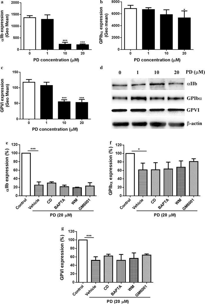
Fig. 7.
Expression of platelet glycoprotein receptors αIIbβ3, GPIbα and GPVI. After PD treatment, the expression of platelet αIIbβ3, GPIbα and GPVI was measured by flow cytometry (mean ± SD, n = 3) (a–c) and western blot (d). Prior to PD treatment, washed platelets were treated with cytochalasin D (CD) (20 μM), BAPTA-AM (BAPTA) (20 μM), wortmannin (WM) (10 μM) or GM6001 (100 μM) followed by measuring the surface expression of platelet αIIbβ3, GPIbα and GPVI by flow cytometry (mean ± SD, n = 3) (e–g). Compared with 0, *P < 0.05; ***P < 0.001

