Fig. 3.

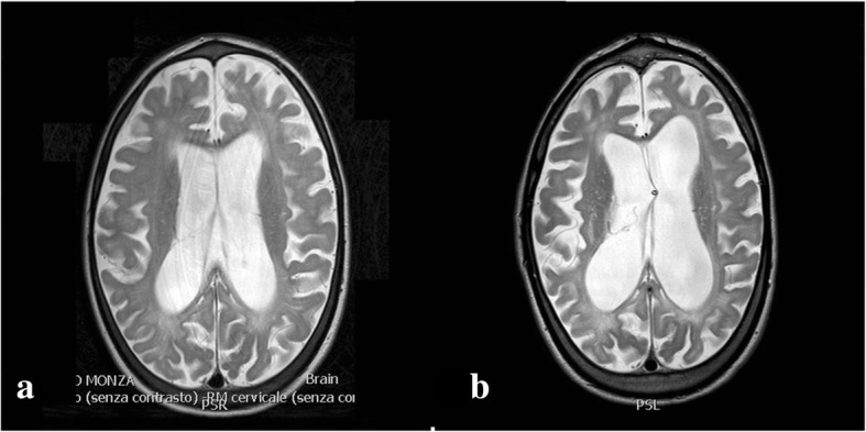
a MRI T2-weighted slices showing ventricular dilatation with transependymal reabsorption. b A ventriculoperitoneal shunt system was placed, with reduction of ventricular volume and disappearance of periventricular imbibition

a MRI T2-weighted slices showing ventricular dilatation with transependymal reabsorption. b A ventriculoperitoneal shunt system was placed, with reduction of ventricular volume and disappearance of periventricular imbibition