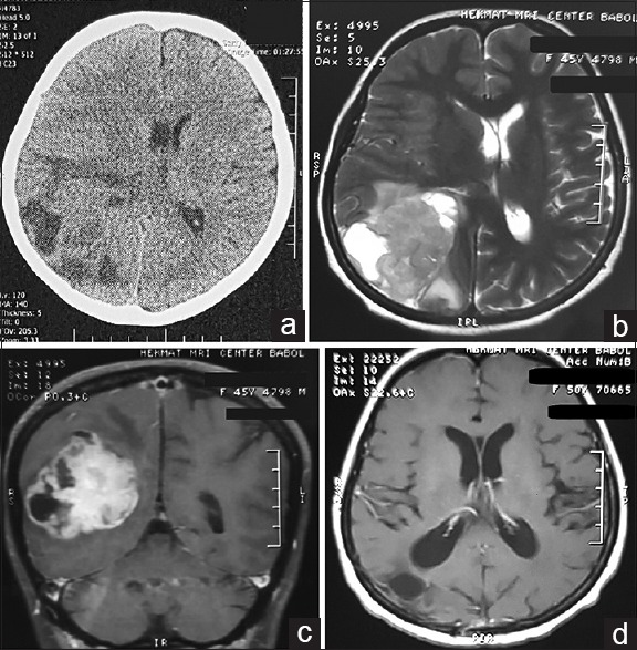
Figure 1.

Preoperative brain computed tomography scan (a) of the patient revealed a large heterogeneous mass at right parieto-occipital region with some vasogenic edema that produces a 0.5 cm midline shift of the brain. On the brain magnetic resonance imaging, we found a solid-cystic hyper-intense mass on T2 sequences (b) that it is brightly enhanced after injection of gadolinium (c). Follow-up brain magnetic resonance imaging with gadolinium revealed no remnant of tumor (d)
