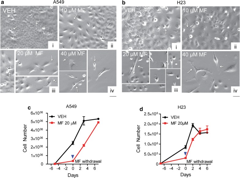
Fig. 3.
Morphological features of NSCLC cells following MF exposure and the consequence of MF withdrawal. In a and b, A549 cells and H23 cells, respectively, were seeded and allowed to attach for 24 h before being treated with the indicated concentrations of MF. Images were taken using phase contrast microscopy following 3 days of treatment. Vehicle-treated cells continued to proliferate (arrows in [i]). Changes in MF-treated cultures included increased cellular size (arrows in [ii]), branching and cytoplasmic extensions with spindle-like morphology (arrows in [iii and iv]. In c and d, A549 cells and H23 cells, respectively, were released of MF treatment on day 0 (blue arrowhead) by replacing MF-containing media with media devoid of MF. At regular intervals, cells were counted using microcapillary cytometry. Negative days are days during which cells were exposed to MF

