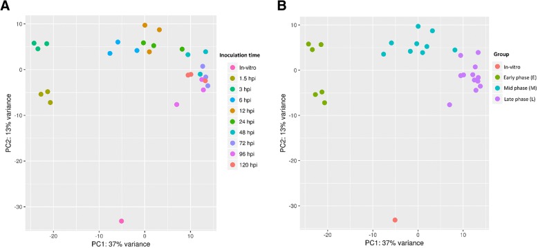
Fig. 2.
Principal component analysis plot (PCA) showing the clustering of VST (variance stabilizing transformation) transcriptomic data. a data points are colored by treatment time point (1.5 HPI, 3 HPI, 6 HPI, 12 HPI, 24 HPI, 48 HPI, 72 HPI, 96 HPI and 120 HPI). b data points are colored by infection phase (in vitro, Early (1.5–3 HPI), Mid (6–24 HPI), Late (48–120 HPI)). HPI, hours post infection

