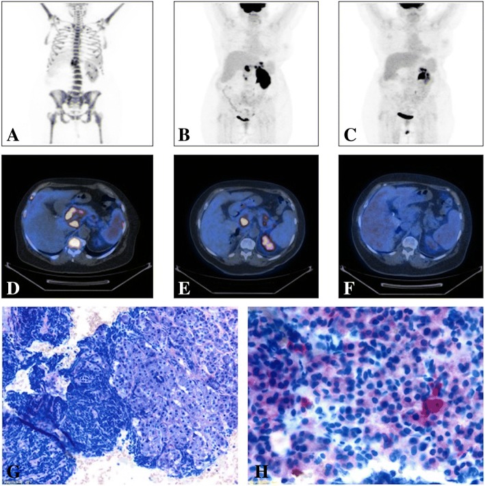
Fig. 1.
Imaging and Histological analysis from Case 1 patient: (a) MIP image (PET-CT scan) before the beginning of IO treatment showing multiple areas of pathological hypermetabolism at patient’s skeleton, spleen and pancreas (SUV max = 32); (b) MIP image (PET-CT scan) after the first course of IO showing the disappearance of the diffuse uptake at patient’s skeleton, a slight reduction of the pancreatic lesion previously reported, with the onset of new areas of pathological uptake within pancreas, gastric fundus and antrum and left renal parenchyma (Deauville V – Progressive Disease); (c) MIP image (PET-CT scan) after the second and last IO course showing a complete metabolic response (Deauville I), with the disappearance of all the previously described lesions; (d) Axial section of a PET-CT scan performed before IO treatments showing pathologic hypermetabolism in pancreas head and body (SUV max = 32, Deauville V), spleen and skeleton (Deauville IV); (e) Axial section of a PET-CT scan performed after the first IO course showing a Progression of the Disease; (f) Axial section of a PET-CT scan performed after the second and last course of IO documenting a Complete Response. (g) Histological image from a Pancreatic echo-endoscopic biopsy showing pancreatic exocrine tubulo-acinar secretory units with massive infiltration represented by B-ALL neoplastic cells (Light Microscopy, Wright-Giemsa); (h) Histological image from a Pancreatic echo-endoscopic biopsy with specific immunostaining showing CD22 slight positivity of B-ALL blasts (Light Microscopy)

