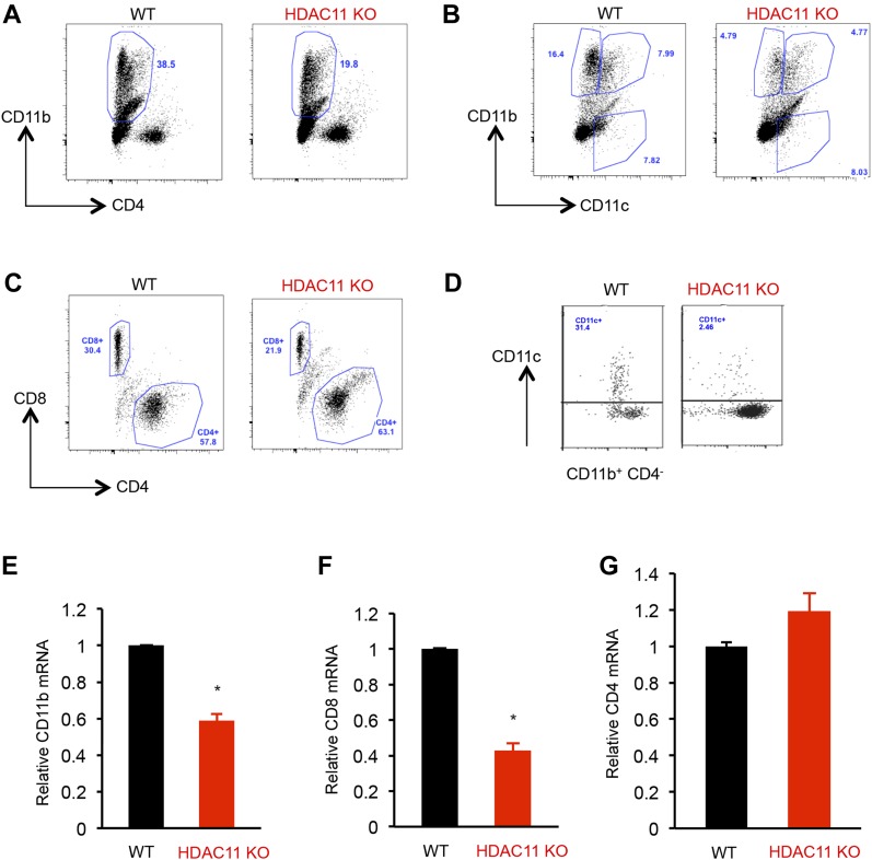
Figure 3. Fewer monocytes and mDCs in HDAC11 KO mice.
(A–C) Flow cytometric analyses of splenocytes isolated from WT and HDAC11 KO mice 12 d after MOG35–55 peptide immunization. Spleen cells were isolated and cultured with 20 μg of MOG35–55 peptide for 72 h and were restimulated with phorbol 12-myristate 13-acetate (PMA) plus ionomycin, with the addition of Brefeldin A for the last 4 h. The cells were stained with antibodies to CD11b-Cy5, CD4-Pacific Blue, CD11c-FITC, or CD8-APC. Analyses were gated on live cells based on live/dead yellow staining. (D) Flow cytometric analyses of mononuclear cells from spinal cords of WT and HDAC11 KO EAE mice, 21 d after MOG35–55 peptide immunization. (E–G) Quantitative polymerase chain reaction (qPCR) analyses of CD11b, CD8, and CD4 mRNA expression in inflammatory cells from spinal cords isolated from WT and HDAC11 KO EAE mice 40 d after MOG35–55 peptide immunization. Data shown are mean ± SD from three independent animals per genotype, and P-values were calculated by t test, *P < 0.05.

