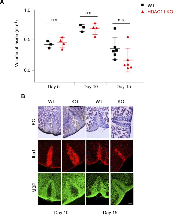
Figure S6. Nonimmune lysolecithin-induced demyelination and remyelination of the spinal cords of WT and HDAC11 KO mice.
10- to 12-wk-old male and female mice were anesthetized with ketamine, xylazine, and acepromazine. Following a level 10 laminectomy, 1.5 μl of 1% solution of LPC (L-α-lysophophatidyl-choline; Sigma-Aldrich) in saline was infused into the dorsal column of spinal cords at 15 μl h −1. Animals were euthanized at 5, 10, and 15 d post injection and their spinal cord segments were extracted and fixed in 4% formalin. (A) Comparison of spinal cords' lesion volumes in WT (day 5, n = 3; day 10, n = 3; day 15, n = 6) and HDAC11 KO (day 5, n = 4; day 10, n = 4; day 15, n = 6) mice. Using Image J, lesion volumes from serial frozen sections (20 μm), stained with Eriochrome (Solochrome) Cyanine (EC), were calculated. Lesion volumes were then matched to the volume of an elliptical cone, V = (π/3)abh, of the lesion midpoint. (B) Myelination was assessed by the staining for nuclear myelin sheath with EC. Spinal cord frozen sections (20 μm) were first fixed in fresh acetone for 5 min, and then stained in 0.2% EC solution for 30 min at RT. Sections were differentiated in 5% iron alum solution for 5–10 min, and were then fully differentiated in 1% borax-ferricyanide solution for 5–10 min. After dehydration with graded ethanol solutions, sections were covered by permanent mounting medium. Fluorescence photomicrographs show staining of activated microglia and macrophages with Iba1 antibodies, and staining of myelin sheath in spinal cords with MBP antibodies (after LPC injections). Scale bar, 100 μm. n.s. represents P > 0.05.

