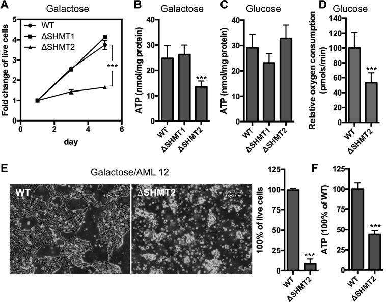
Figure 2. Effects on mitochondrial respiration by the deletion of serine catabolic enzymes in 293A and AML12 cells.
(A) Measurement of the cell proliferation of the WT, ΔSHMT1, and ΔSHMT2 293A cells in the DMEM-based galactose media. (B, C) Measurement of the intracellular ATP levels in the WT, ΔSHMT1, and ΔSHMT2 293A cells that were grown in the galactose (B) or glucose (C) media for 24 h. (D) Measurement of the basal oxygen consumption rates of the WT and ΔSHMT2 293A cells that were grown in the glucose media. (E) Phase-contrast images illustrating the WT and ΔSHMT2 AML12 cells that were grown in the galactose media for 72 h. The percentages of the live cells were plotted on the right. (F) Measurement of the intracellular ATP levels of the WT and ΔSHMT2 AML12 cells that were grown in the galactose media for 72 h. ***P < 0.001 (t test). Data are presented as mean ± SD. n = 5 for (A); n = 4 for (B–E); and n = 6 for (F).

