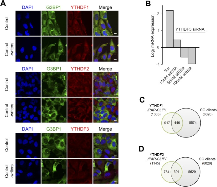
Figure S4. Localization and clients of the YTHDF readers.
(A) Cytoplasmic localization of YTHDF1, YTHDF2, and YTHDF3 in control, unstressed cells and by silenced “writers” (METTL3, METTL14, and WTAP). The “readers” were detected with Alexa568-conjugated secondary antibody (red). Scale bar, 10 μm. (B) qRT–PCR following siRNA knockdown of YTHDF3 for 48 h in U2OS-G3BP1 cells. The YTHDF3 expression was normalized to the expression of β-actin mRNA. Scr denotes scrambled siRNA and accounts for unspecific effects. (C) mRNA clients of YTHDF1 identified by PAR-CLIP in Wang et al (2015) and compared with the SG transcripts identified in this study. P = 0.006, hypergeometric test. (D) mRNA clients of YTHDF2 identified by PAR-CLIP in Wang et al (2014) and compared with the SG transcripts identified in this study. P = 3.9 × 10⁻⁴, hypergeometric test.
Source data are available for this figure.

