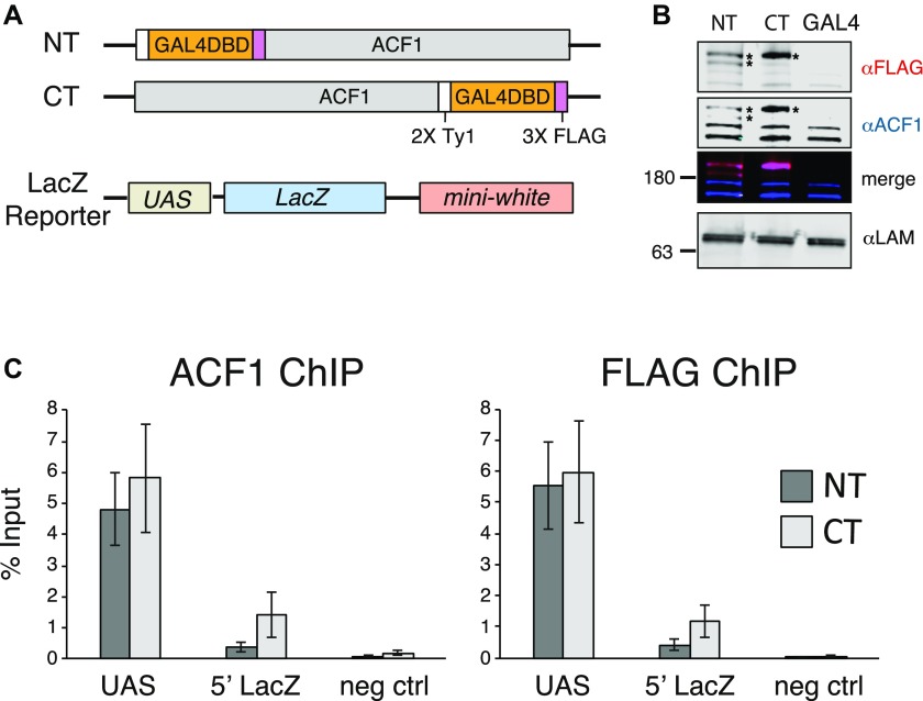
Figure 1. ACF can be tethered to a reporter locus through GAL4-DBD.
(A) Schematic illustration of the transgenes used for testing the effects of ACF1 recruitment. ACF1 is fused to the GAL4 DNA-binding domain (GAL4DBD) at either the N-terminus or the C-terminus. A transgene containing the GAL4DBD alone (GAL4) is used as a negative control. The reporter transgene contains five UASGal4 (UAS) 5′ of LacZ and mini-white genes. (B) Western blot detection of ACF1 in an embryo nuclear extract (0–16 h AEL). Endogenous and fusion proteins were detected with a specific ACF1 antibody (blue channel); the ACF1-GAL4 fusions are FLAG-tagged and detected with an anti-FLAG antibody (magenta channel). Asterisks indicate the expressed transgenic ACF1-GAL4 fusions. Embryos containing a transgene coding for the GAL4DBD alone (GAL4) are included as a negative control. Lamin serves as a loading control. (C) ChIP-qPCR monitors the recruitment of ACF1 to UAS in 0- to 12-h embryos. The immunoprecipitation was conducted using ACF1 and FLAG antibodies. “UAS” and “5′ LacZ” denote the regions amplified by qPCR. Bars denote average % Input enrichment (n = 3 biological replicates) ± SEM. “neg ctrl” represents a negative control locus (encompassing the Spt4 gene).

