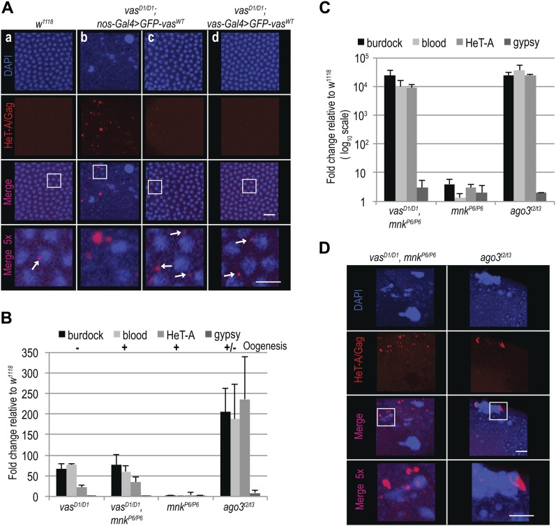
Figure 3. Loss of Chk2 DNA damage signaling does not restore embryogenesis in vas mutant flies.
(A) Immunohistochemical detection of HeT-A/Gag protein in WT (w1118; a), vasD1/D1; nos-Gal4>GFP-vasWT (b and c), and vasD1/D1; vas-Gal4>GFP-vasWT (d) stage 5 embryos. Arrows indicate WT localization of HeT-A/Gag. Staining of the whole embryos is presented in Fig S3A. Scale bars indicate 10 and 5 μm (5× magnification). (B) qPCR analysis of LTR transposons burdock, blood, and gypsy and non-LTR transposon HeT-A RNAs in ovaries from vasD1/D1 single and vasD1/D1, mnkP6/P6 double mutant flies, and mnkP6/P6 and agot2/t3 mutant flies. The expression level of transposons in WT (w1118) was set to one and normalized to rp49 mRNA in individual experiments. Error bars represent the standard deviation among three biological replicates. Oogenesis completion is indicated with + and −. (C) qPCR analysis of LTR transposons burdock, blood, and gypsy and non-LTR transposon HeT-A RNAs in early embryos produced by vasD1/D1, mnkP6/P6 double mutant, and mnkP6/P6 and agot2/t3 single mutant flies. The expression level of transposons in WT (w1118) was set to one and normalized to 18S rRNA in individual experiments. Error bars represent the SD among three biological replicates. (D) Immunohistochemical detection of HeT-A/Gag protein in stage 5 embryos from vasD1/D1, mnkP6/P6 double mutant and agot2/t3 single mutant flies. Staining of the whole embryos is presented in Fig S4D. Scale bars indicate 10 and 5 μm (5× magnification).

