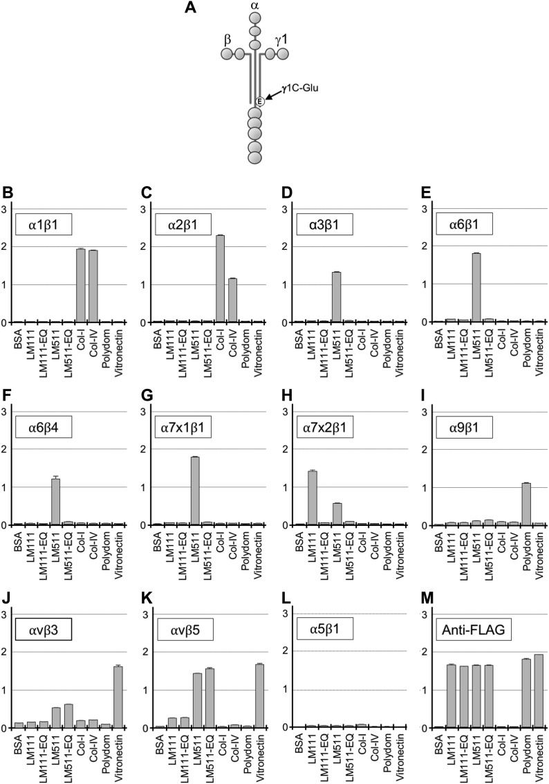
Figure 1. Binding of recombinant laminin-111, laminin-511, and their EQ mutants to integrins.
(A) Schematic diagram of a full-length laminin. The Glu (E) residue in the C-terminal region of the γ1 chain critical for integrin binding is indicated. (B–L) Binding of recombinant integrin α1β1 (B), α2β1 (C), α3β1 (D), α6β1 (E), α6β4 (F), α7x1β1 (G), α7x2β1 (H), α9β1 (I), αvβ3 (J), αvβ5 (K), and α5β1 (L) to immobilized laminins (LMs), type-I and type-IV collagens, polydom, and vitronectin. (M) Anti-FLAG antibody binding for quantification of immobilized recombinant proteins. Vertical axes represent absorbance at 490 nm which indicates integrin binding. Data represent means ± SD of triplicate assays.

