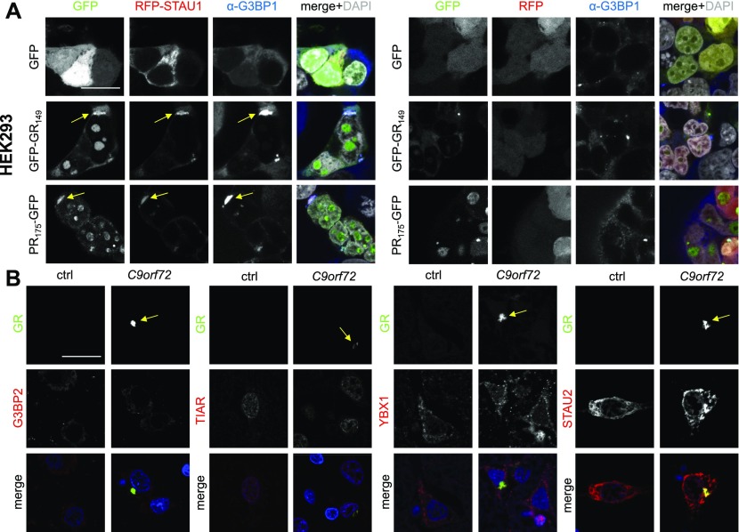
Figure 4. Cytoplasmic poly-GR/PR inclusions resemble stress granules in vitro.
Immunofluorescence of stress granule markers in HEK293 cells and patient brain. DAPI visualizes nuclei. Single confocal planes were taken. Scale bar depicts 20 μm. (A) Co-localization of poly-GR/PR with the stress granule marker G3BP1 in HEK293 cells co-transfected with RFP-STAU1 or RFP control and DPR-GFP or GFP control. Left three columns show individual channels as indicated. The fourth columns show merge with additional nuclear DAPI staining in white. Arrows indicate cytoplasmic inclusions co-labeled with G3BP1. (B) Immunofluorescence of frontal cortex of a C9orf72 patient and a healthy control case to analyze co-localization of poly-GR with stress granule components TIAR, G3BP2, YBX1, and STAU2. Arrows indicate poly-GR aggregates.
Source data are available for this figure.

