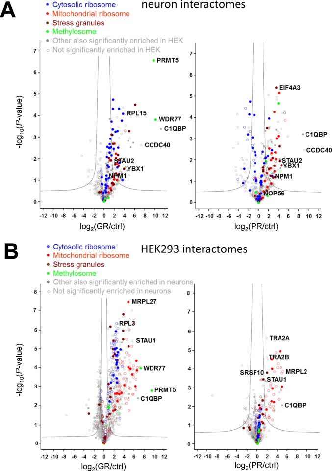
Figure S2. Poly-GR and poly-PR interact with overlapping proteins in primary neurons and in HEK293 cells.
Quantitative proteomics of GFP immunoprecipitations from HEK293 cells and primary rat neurons transfected or transduced with GFP, GFP-(GR)149, or (PR)175-GFP. (A, B) Volcano plots of interactomes from neurons (A) and HEK293 cells (B). The data for all proteins are plotted as log2-fold change versus the −log10 of the P-value. Gray line indicates significance cutoff. Enriched protein families are color coded: cytoplasmic ribosome (blue), mitochondrial ribosome (red), stress granules (brown) (Jain et al, 2016), and methylosome (green). The top enriched proteins (sorted by fold-change) and the proteins analyzed in this study are labeled with gene names. Filled circles indicated that the protein was significantly altered in the other cell type.

