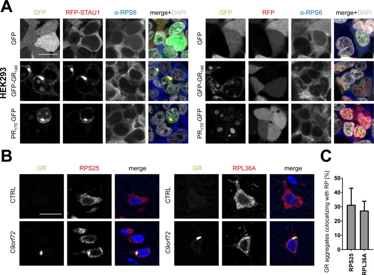
Figure S4. Poly-GR mainly co-localizes with nucleolar ribosomes.
(A) HEK293 cells were co-transfected with GFP, GFP-(GR)149, or (PR)175-GFP expression vectors and RFP-STAU1 as in Fig 3A and endogenous RPS6 detected by immunofluorescence. Note that the large cytoplasmic poly-GR/PR granules induced by STAU1 expression are not clearly stained with the ribosomal subunit compared with the cytoplasm. Merge shows additionally DAPI in gray. Single confocal planes are shown. Scale bar denotes 20 μm. (B) Frontal cortex of a C9orf72 patient and a control brain were co-stained for poly-GR and ribosomal proteins as in Fig 5A. RPS25 and RPL36A are partially sequestered into poly-GR inclusions. DAPI visualizes nuclei. Single confocal planes are shown. Scale bar depicts 20 μm. (C) Quantification of GR aggregates co-localizing with ribosomal proteins (n = 3 cortical sections with 100 GR aggregates counted each from two different patients; mean ± SEM is shown).

