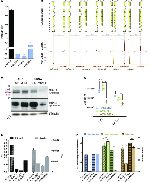
Figure 3. The exclusion of exon 7 through the use of Antisense oligonucleotides reduces cell proliferation.
(A) MBNL1 ex7 PSI change upon AON transfections (100 nM), 48 h posttransfection. Data are representative of two independent experiments. (B) FLA-PCR visualization of MBNL1 cDNA isoforms upon AON #592 transfection in PC3 cells (100 nM, at 48 h posttransfection). On the X axis is represented the nucleotide size and on the Y axis the fluorescence intensity of the amplicon. Data are representative of multiple independent experiments. (C) Western blot of PC3 cells transfected with AON (100 nM) and siRNA (25 nM) directed against MBNL1. Cells were collected 48 h posttransfection. (D) PC3 and LnCap absolute cell number at 48 h posttransfection with AON SCR, MBNL1 ex7 (100 nM), or not transfected (no liposome-based reagent, no AON). Statistics performed with two-tailed, parametric, paired t test. (E) MBNL1 ex7 PSI change and relative cell viability in PC3 cells. 100 nM of AON after 48 h. Cell viability quantified with TiterGlo luminescence assay. (F) Ex7 PSI (blue bars), overall MBNL1 relative mRNA expression (green bars) and relative cell count of PC3 cells transfected with AON SCR, AON MBNL1 (100 nM), siRNA Scrambled, and siRNA MBNL1 (25 nM).

