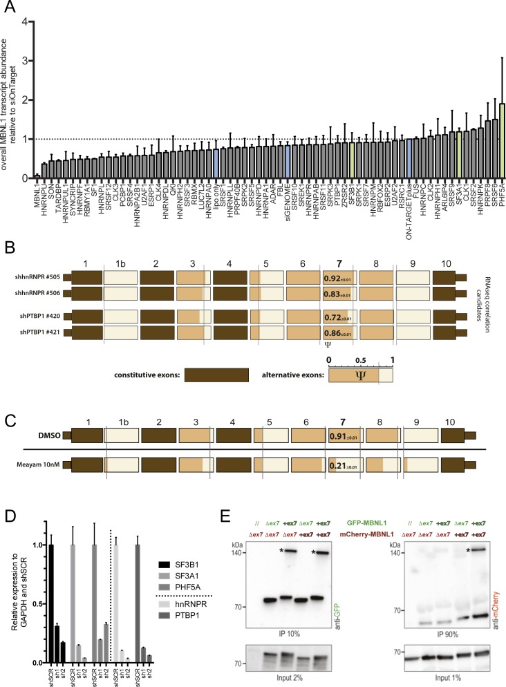
Figure S2. Splicing factor's siRNA screening and MBNL1 co-IP.
(A) Overall MBNL1 mRNA transcript abundance (including and excluding ex7) normalized for GAPDH and siON-TARGET plus control. (B) shRNA validation of MBNL1 ex7 modulator's negative controls in PC3 cells. The PSI of every alternative exon is shown in orange. Constitutive exons are colored in brown. For ex7, the values of the PSI are written in black. Gray line shows the shSCR value. Three biological replicates for each shRNA are shown. (C) Effects on MBNL1 AS of meayamycin (10 nM) in PC3 cells after 20 h treatment. All data represented are obtained from two independent biological replicates. (D) Relative mRNA expression of SF3B1, SF3A1, PHF5A, and the control genes hnRNPR and PTBP1 upon shRNA-mediated knockdown. (E) Co-immunoprecipitation with anti-GFP beads. Western blots obtained with antibodies against GFP (left) or mCherry (right). The co-overexpression combination in Cos7 cells of the isoforms C (with ex7) and H (lacking ex7) fused with GFP or mCherry is shown above the graph. Asterisks indicate plausible dimers of MBNL1 proteins. Data shown are representative of three independent biological replicates.

