
Figure S1. Western blot of Aβ42-specific antibody compared with 4G8 antibody.
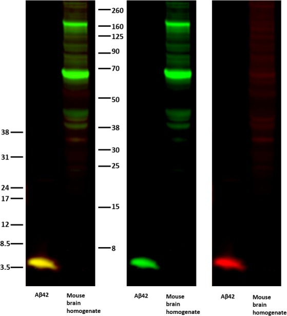
WB of purified Aβ42 and mouse brain homogenate samples were incubated with the Aβ42-specific antibody (anti-AβC42, red) and an antibody recognizing the Aβ sequence within APP (4G8, green). The merged channel is shown to the left, and the individual channels are shown in the middle and to the right. The molecular weight is shown by an Odyssey Protein Molecular Weight Marker (LI-COR) on the right hand side and low-range Amersham Rainbow Marker (RPN 755E; GE Healthcare) on the left hand side.