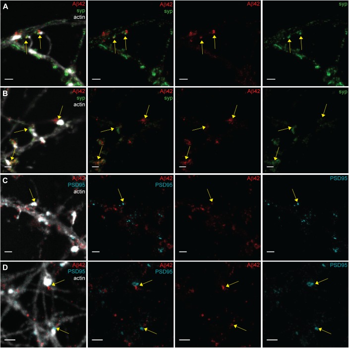
Figure S3. STED images of Aβ42 focusing on the pre- or postsynaptic region in hippocampal neurons.
(A, B) Zoomed STED images of synapses showing Aβ42 (red) and the presynaptic marker synaptophysin (green) combined with actin in a confocal channel (light grey) in the left panel. The staining of synaptophysin and Aβ42 at presynaptic boutons connected to the postsynaptic spines are shown by the yellow arrows. Scale bars: 1 μm. (C, D) Zoomed STED images of synapses showing Aβ42 (red) and the presynaptic marker PSD95 (cyan) combined with actin in a confocal channel (light grey) in the left panel. The staining of PSD95 at postsynaptic spines and Aβ42 at presynaptic boutons is shown (yellow arrows). Scale bars: 1 μm.

