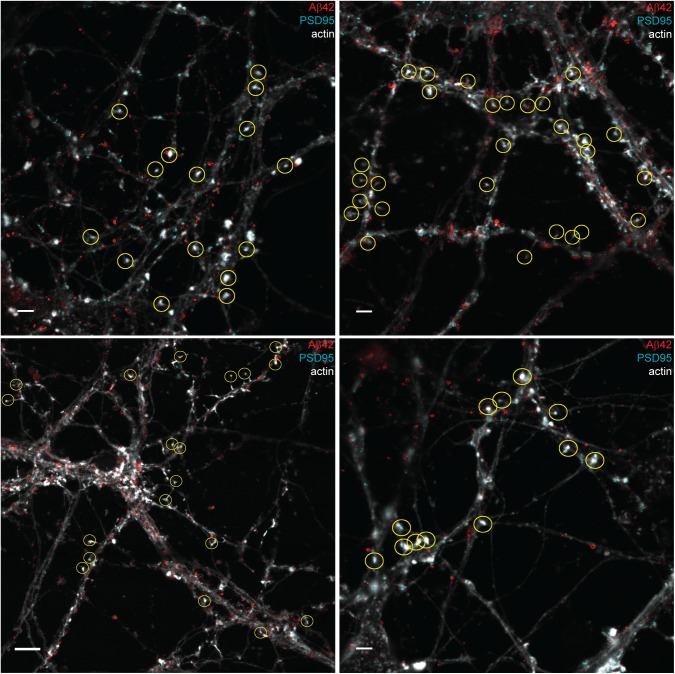
Figure S7. Synapses included in quantification of Aβ42 staining in postsynapses in hippocampal neurons.
STED images stained with Aβ42 (red) and PSD95 (cyan) combined with an actin (light grey) confocal channel. The yellow circles show synapses selected for quantification: 73 in total. Bottom left scale bar: 5 μm. Other scale bars: 2 μm.

