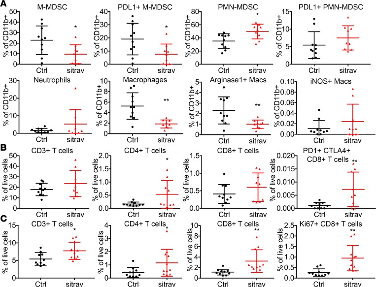
Figure 5. Sitravatinib alters the immune landscape of KLN205 tumors to favor immune checkpoint blockade.
Flow cytometry of tumor-associated myeloid (A) and lymphoid cells (B) from mice bearing KLN205 tumors treated for 6 days with sitravatinib (sitra, n = 9–10/group). Monocytic myeloid-derived suppressor cells (M-MDSCs; CD11b+Ly6G–Ly6C+), PD-L1+ M-MDSCs, polymorphonuclear myeloid-derived suppressor cells (PMN-MDSCs; CD11b+Ly6G+Ly6C+), PD-L1+ PMN-MDSC, neutrophils (CD11b+Ly6G+Ly6C–), macrophages (CD11b+Ly6G–Ly6C–F4/80+CD11c+MHCII+), Arg1+ macrophages (Macs), iNOS+ macrophages, CD3+ T cells, CD4+ T cells, CD8+ T cells, and PD-1+CTLA4+CD8+ T cells were analyzed. *P < 0.05, **P < 0.01 vs. control (Ctrl) by t test. (C) Flow cytometry of splenocytes from mice bearing KLN205 tumors treated with sitravatinib for 6 days (n = 9–10/group). CD3+ T cells, CD4+ T cells, CD8+ T cells, and Ki67+ CD8+ T cells were analyzed. *P < 0.05, **P < 0.01 vs. control by t test.

