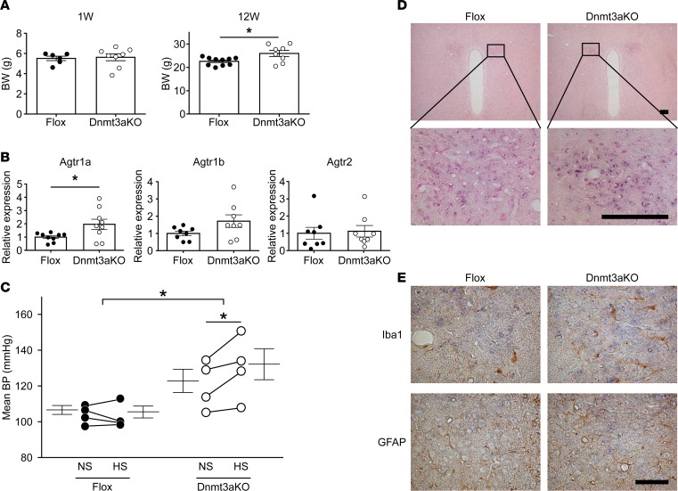
Figure 5. Salt-sensitive hypertension in offspring of hypothalamic neuron–specific Dnmt3a-KO mice without dexamethasone (Dex) treatment during pregnancy.
(A) BW at weeks 1 and 12 in the flox mice (n = 6 and 10, respectively) and hypothalamic neuron-specific Dnmt3a-KO mice (n = 8 and 8, respectively). Filled circle, Dex-untreated flox mice; open circle, Dex-untreated Dnmt3a-KO mice. (B) Real-time PCR of Agtr1a (n = 9), Agtr1b (n = 8), and Agtr2 (n = 8) mRNA in PVN of flox and Dnmt3a-KO mice. Filled circle, Dex-untreated flox mice; open circle, Dex-untreated Dnmt3a-KO mice. (C) Mean BP by radiotelemetry before and after 1 week of HS in Dex-untreated flox mice (n = 4, left) and Dex-untreated Dnmt3a-KO mice (n = 4, right). Filled circle, Dex-untreated flox mice; open circles, Dex-untreated Dnmt3a-KO mice. In A and B, *P < 0.05 versus Dex-untreated flox mice (t test); in C, *P < 0.05 versus HS-treated flox mice or NS-treated Dnmt3a-KO mice (2-way repeated ANOVA, Bonferroni post hoc text). (D) In situ hybridization of Agtr1a mRNA in the PVN of Sim1-Cre Dnmt3a-KO (right) and flox mice (left) (upper panels, low power; lower panels, high power). Hybridization using an antisense probe indicates expression of Agtr1a mRNA (purple, lower panel). Hybridization using the sense probe yielded no detectable signals in the PVN (data not shown). Scale bar: 100 μm. (E) Upper panels show staining for Iba1, a marker of glia cells (brown), and lower panels show GFAP, a marker of astrocytes (brown). Agtr1a (blue) colocalized with neither Iba1 nor GFAP, suggesting that it is expressed mainly in neuronal cells. The expression pattern was not affected by Sim1-Cre Dnmt3a–KO. Scale bars: 50 μm.

