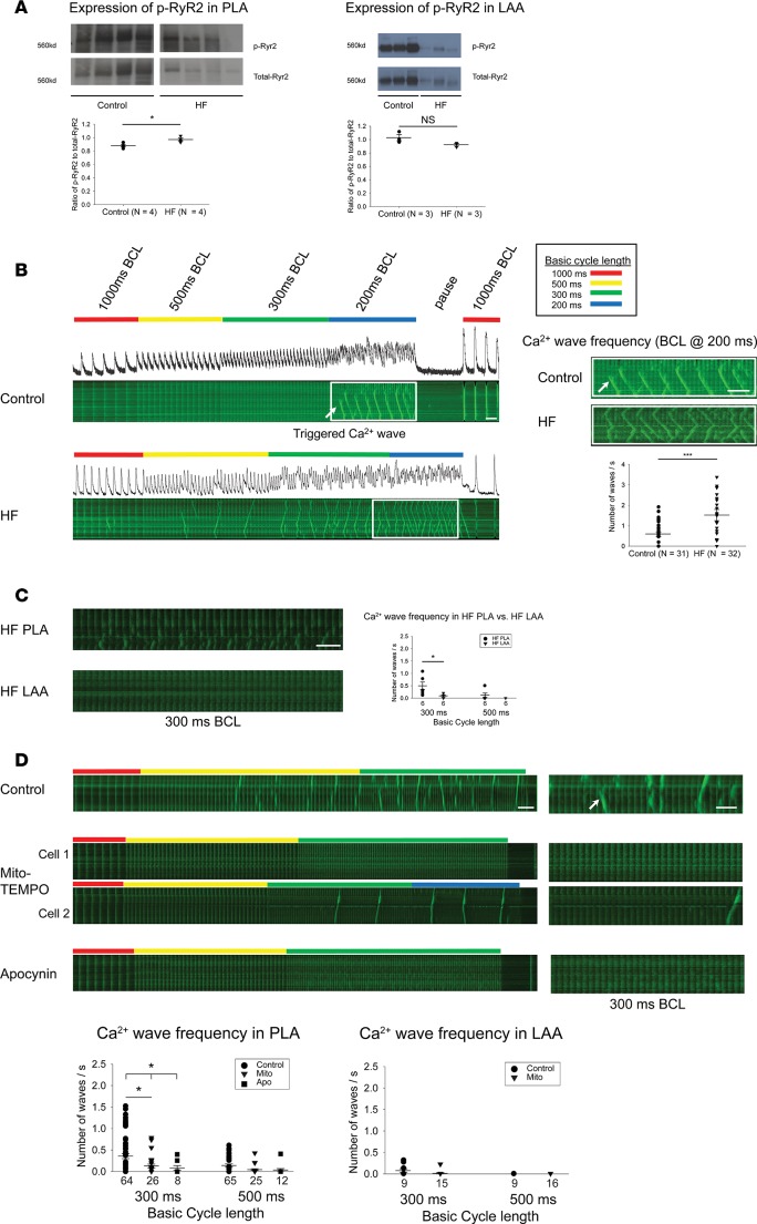
Figure 3. Increased expression of CaMKII-p-RyR2 (S2814) in HF PLA and sensitivity of triggered Ca2+ waves in PLA myocytes to acute ROS inhibition.
(A) Representative immunoblot and densitometric measurements of CaMKII-p-RyR2 (S2814) (normalized to native RyR2) from control and HF in PLA (left) and LAA (right). (B) HF atrial myocytes showed higher incidence of triggered Ca2+ waves, compared with control. (C) Higher incidence of triggered Ca2+ waves in HF PLA myocytes, compared with HF LAA myocytes at 300 ms BCL. (D) Ca2+ imaging showed attenuation of incidence of triggered Ca2+ waves at 500 ms and 300 ms BCL in the presence of mito-TEMPO and apocynin in HF PLA myocytes, but not in HF LAA myocytes. Time bar: 1 second. The myocytes for these experiments were obtained from 4 HF dogs, and the number of myocytes for each experimental condition is given in each figure panel. Data are represented as mean ± SEM. *P < 0.05, ***P < 0.001. (A and B) Independent t test. (C) Paired t test. (D) Main effect of ANOVA (P = 0.017 at 300 ms, 0.053 at 500ms), post hoc t tests with each cycle with Bonferroni correction. HF, heart failure; PLA, posterior left atrium; LAA, left atrial appendage. See complete unedited blots in the supplemental material.

