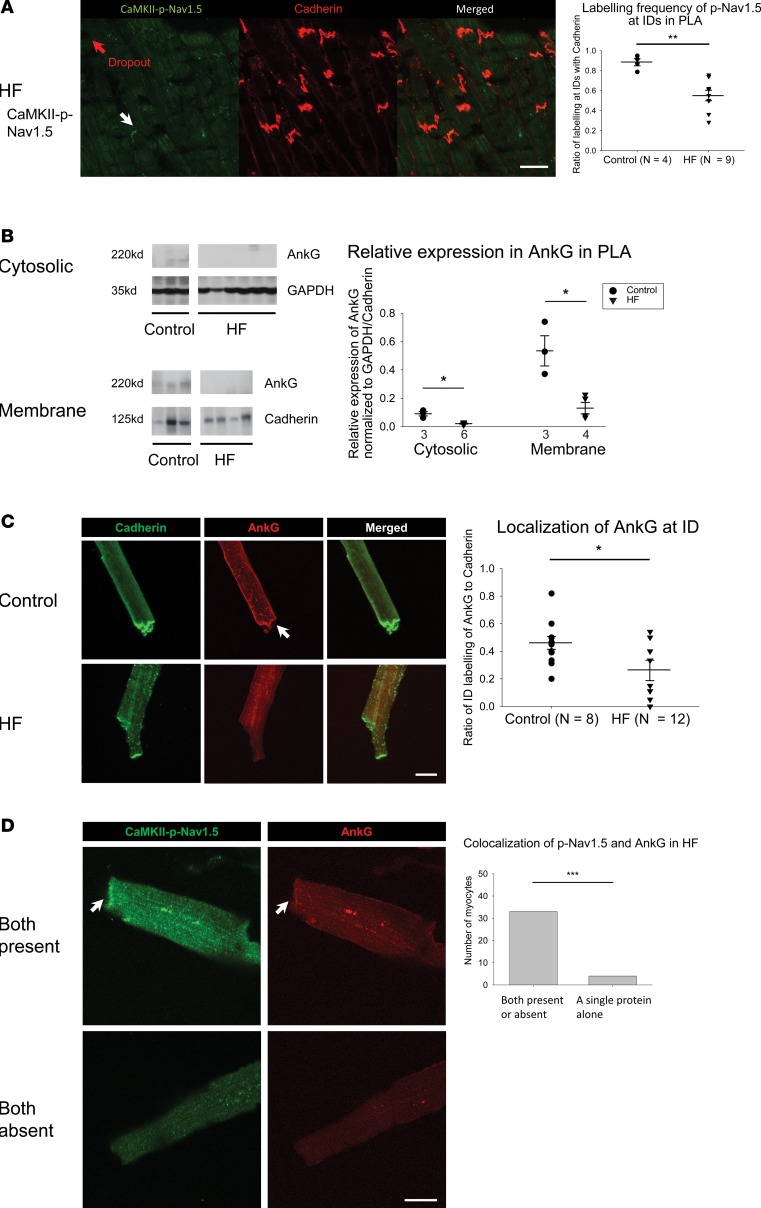
Figure 6. Decrease in expression and ID localization of AnkG is responsible for dropout of CaMKII-p-Nav1.5 (S571) in HF PLA.
(A) In HF PLA, there was a dropout of CaMKII-p-Nav1.5 (S571) at certain ID (red arrow), where cadherin labeling was still intact at those ID (left). Quantification of myocytes with ID labeling of CaMKII-p-Nav1.5 (S571) in control and HF PLA (right). (B) Representative immunoblot and densitometric measurements of AnkG (normalized to GAPDH and cadherin) in cytosolic and membrane fractions, respectively, from control and HF PLA. (C) ID localization of AnkG (red) was reduced in isolated HF myocytes, while labeling of cadherin (green) seemed similar in control and HF myocytes. (D) Colocalization of CaMKII-p-Nav1.5 (S571) and AnkG at the ID (white arrows). Scale bar: 40 μm. Data are represented as mean ± SEM. *P < 0.05, **P < 0.01, ***P < 0.001. (A and B) Independent t test. (C) Generalized linear model, logit link comparing rates if ID localization of AnkG. (D) χ2 test. HF, heart failure; PLA, posterior left atrium; ID, intercalated disc. See complete unedited blots in the supplemental material.

