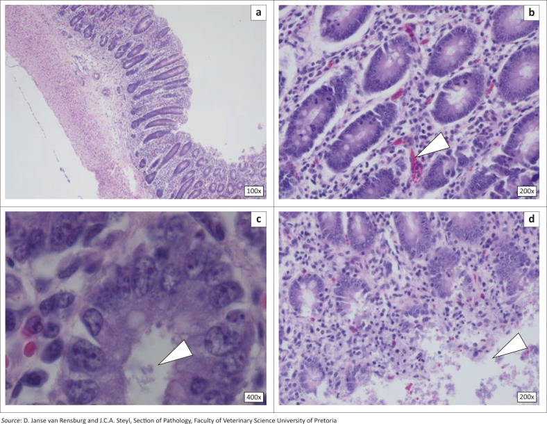
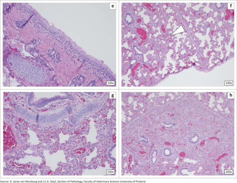
FIGURE 1.
Histopathology of the intestinal and respiratory system tissue from the studied animals, stained with haematoxylin–eosin. (a) Overview of a transverse section of intestine; (b) Intestinal lumen, with normal lymphocytes and plasma cells in the lamina, mild congestion of enterocytes and lymphoid cells (arrow) and normal mucosal cells; (c) Lumen filled with bacteria and debris (arrow), normal enterocytes and goblet cells; (d) Section of the intestine, showing normal enterocytes, congestion, mild haemorrhage and autolytic cells (arrow); (e) Transverse section of the pig trachea showing the respiratory epithelium, blood vessels and hyaline cartilage; (f) Interstitial alveolar wall thickening (arrow); (g) Interstitial pneumonia and congestion; (h) Consolidated lung tissue with numerous bronchioles and congestion.


